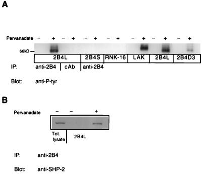
Figure 7.
2B4L tyrosine phosphorylation and SHP-2 association. (A) Approximately 2 × 10⁷ cells of various RNK-16 transfectants or C57/Bl6 LAKs were incubated at 37°C for 10 min in the presence or absence of 1 mM pervanadate. Cells were lysed and immunoprecipitated with 5 μg of anti-2B4 antibody or control antibody (cAb) as indicated. Immunoprecipitates were resolved by nonreducing SDS/PAGE and immunoblotted with horseradish peroxidase-conjugated anti-phosphotyrosine antibody and analyzed by enhanced chemiluminescence. Bands corresponding to 2B4 are located near the 66-kDa marker. (B) Approximately 1 × 10⁸ cells of 2B4L expressing RNK-16 cells were incubated with or without pervanadate as described above. Cells were then lysed and subjected to immunoprecipitation with anti-2B4 antibody. Immunoprecipitates were then resolved by nondenaturing SDS/PAGE and subjected to immunoblotting with anti-SHP-2 antibody followed by horseradish peroxidase-conjugated secondary antibody and analyzed by enhanced chemiluminescence. The lane marked total lysate represents 1 × 10⁶ cell equivalents loaded onto the gel.

