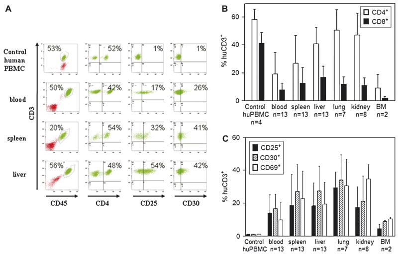
Figure 4.
Human T cell infiltration and phenotype in tissues of NOD/SCID-β2mnull mice that develop lethal GVHD. NOD/SCID-β2mnull mice (n=34) were sublethally irradiated with 250cGy of TBI and injected r.o. with 10 × 106 naive purified huT cells. When possible, moribund animals with X-GVHD were euthanized and portions of blood, spleen, liver, lung, kidney and bone marrow (BM) were analyzed by flow cytometry. (A) Representative FACS analysis of blood, spleen, and liver harvested from a NOD/SCID-β2mnull mouse that developed lethal X-GVHD. In all FACS analyses, normal human PBMCs were used as controls to establish consistent gating criteria. (B) Human T cell subset composition. The percentage of human CD45+CD3+ T cells in the indicated tissues that expressed CD4 or CD8 was determined by flow cytometry. (C) Expression of T cell activation markers. The percentage of human CD45+CD3+ T cells in the indicated tissues that expressed CD25, CD30, or CD69 was determined by flow cytometry

