
Fig. 4.
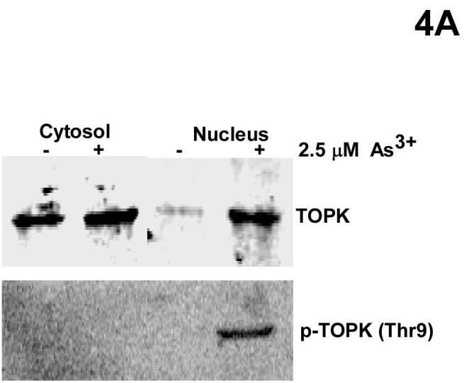

TOPK and histone H2AX co-localize in the nucleus. A, cytosolic and nuclear localization of total and phosphorylated TOPK in RPMI7951 cells after 24 h treatment with 2.5 μM As3+. Cytosolic and nuclear proteins were extracted and separated by 10% SDS-PAGE followed by Western blot analysis with specific antibodies against phosphorylated and nonphosphorylated TOPK. B, nuclear co-localization of phosphorylated TOPK and phosphorylated H2AX. RPMI7951 cells were or were not treated with 2.5 μM As3+ for 24 h. TOPK was visualized under a fluorescence microscope using an FITC-specific antibody (upper panels) and histone H2AX was visualized (middle panels) using a Texas Red-conjugated antibody. Pictures of As3+ treated (right panels) and untreated (left panel) cells represent exactly the same region for each, respectively, allowing the lower panels to demonstrate the merged staining result (x630). Scale bar, 25 μm. These data are representative of at least three independent experiments.