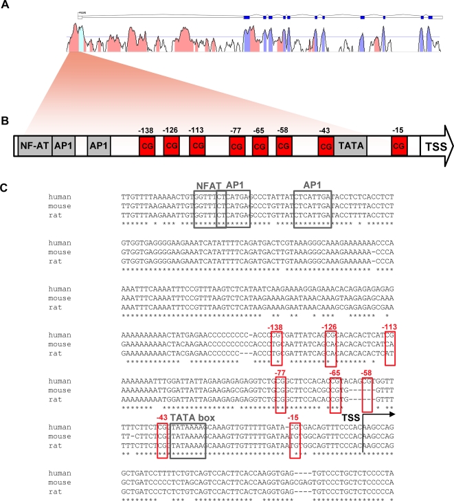
Figure 2. Identification of cross-species conserved FOXP3 promoter elements.
(A) FOXP3 transcript and m-Vista alignment showing conservation between human and mouse genomic sequences. Dark blue boxes display exons, outlined boxes are UTR's. Conserved regions are in red and conserved regions corresponding to exons are in light blue. (B) Schematic view of the conserved region upstream of the transcription start site indicating the location of promoter elements and CpG dinucleotides. (C) Clustal W alignment of human, mouse and rat genomic sequences showing a detailed view of the conservation at the FOXP3 core promoter. The broken arrow shows the position of the human transcription start site. The location of the TATA box is indicated by a black box. Red boxes indicate the CpG dinucleotides analyzed in this study. Transcription factor binding sites are marked with grey boxes.

