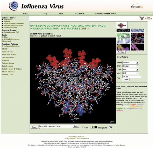
Figure 4.
3D Protein structure visualization. A ball-and-stick representation of the RNA-binding domain of the NS1 protein dimer from the A/Udorn/307/1972 (PDB ID 1NS1) is shown. RNA-binding residues aa38 and aa41 are highlighted in red, while the single amino acid difference between DkXi22 and DkXi35 at aa66 is highlighted in blue. RNA binds parallel to the plane on top of the red RNA-binding residues.

