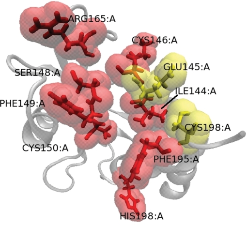
Figure 4.
View of numb protein phosphotyrosine binding (PTB) domain. Red and yellow residues are experimental hot spots. Red residues are correctly predicted by HotSprint. Left and right figures present the results for the prediction of hot spots using pScore and pScore + ASA, respectively. VMD (29) is used to graphically represent the protein.

