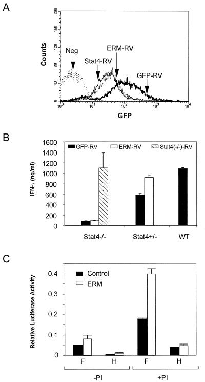
Figure 5.
Effects of ERM expression on IFN-γ production in Stat4-deficient and Stat4-herterozygous T cells. (A) Stat4-deficient, Stat4-heterozygous, and wild-type DO11.10 TCR-transgenic T cells were activated in vitro by using OVA/APCs and infected on day 1 after primary activation with either empty vector (GFP-RV), Stat4-expresing retrovirus (Stat4-RV), or ERM-expressing retrovirus (ERM-RV). Infected cells were purified by cell sorting on day 7 for GFP (FL1) and CD4 expression by using anti-mouse-CD4-Phycoerythrin (PharMingen). Sorted GFP+/CD4+ T cells were reactivated with OVA, APCs, and IL-12 (see Methods), and stable transfection was confirmed by GFP expression by FACS analysis 3 days later. Data shown are single color histograms of the indicated sorted transfectant populations. The negative control (Neg) is an uninfected, Stat4-deficient DO11.10 T cell population activated concurrently under the same conditions. (B) Retrovirally infected T cells from the indicated populations described in A were harvested, washed, and restimulated at 1.25 × 10⁵/ml with 0.3 μM of OVA and irradiated BALB/c splenocytes as APCs. Supernatants were harvested after 48 hr, and IFN-γ production was determined by ELISA. Similar results were obtained in three similar independent experiments. (C) Luciferase reporter assays were performed as described in Methods by using the IFN-γ promoter constructs F and H. Transfected cells were treated with phorbol 12-myristate 13-acetate and ionomycin (PI+) or left untreated (PI−) as described (25). Data are presented as relative light units after normalization for transfection efficiency by using CMV-Renilla luciferase.

