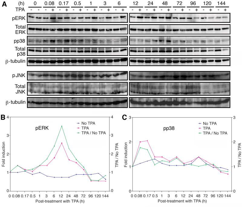
Fig. 1.
Constitutive activation of the ERK, JNK and p38 MAPK pathways in uninduced BCBL-1 cells, and transient enhanced activation of the ERK but not JNK and p38 pathways by TPA. (A) Western-blotting with specific antibodies to detect the phosphorylated forms of ERK (first set of panels), p38 (third set of panels) and JNK (sixth set of panels), and total ERK (second set of panels), p38 (fourth set of panels) and JNK (seventh set of panels) in cells induced with TPA for different lengths of time (0-144 h). An anti-β-tubulin antibody was used to normalize the sample loading (fifth and eighth panels). The ERK and p38 blots were run with one set of samples while the JNK blots were run with another set of samples. (B) Quantification of pERK levels in mock- and TPA-induced cells. (C) Quantification of pp38 levels in mock- and TPA-induced cells.

