
FIGURE 4.
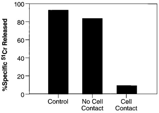
Suppression of CD8+ T cell function requires cell-cell contact. A suspension of three spleens from BALB/c mice infected 6 days earlier with 5 × 10⁶ PFU/mouse of IFN-γ-rVV was incubated with a β-gal peptide either alone (control) or together with splenocytes of mice similarly infected with IL-2-rVV (1:1). The latter were either admixed in the same well (cell contact) or separated by a semipermeable membrane (no cell contact). After 6 days, CTL activity in the cultures was assayed against the same panel of target cells used in previous experiments. For simplicity, only the cytolytic activity against CT26.CL25 at an E:T ratio of 10:1 is shown. Control wells containing 1:1 mixture of splenocytes from IFN-γ-rVV-infected mice and normal mice did not show any difference from control culture whether they were admixed or separated by the insert membrane (not shown).