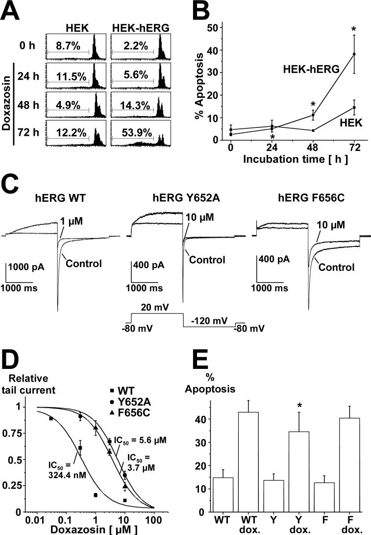
Fig. 2.
A Flow cytometric analysis illustrating time-dependence of pro-apoptotic effects of doxazosin. Cells were treated with 30 μM doxazosin and analyzed for subdiploid DNA content as hallmark for apoptosis using the assay described by Nicoletti et al. (1991). One representative FACS result is shown. B Mean values (± S.E.M) obtained from three experiments (* indicates p < 0.05 versus drug-free conditions at 0 h). Statistical significance was calculated using Student's t tests. C Attenuation of doxazosin block by pore mutations demonstrates direct doxazosin binding to hERG channels. Original current traces are shown, illustrating the effects of doxazosin on wild type or mutant Y652A and F656C currents (1 and 10 μM doxazosin, respectively). D Concentration-response relationships for doxazosin blockade of hERG constructs transiently expressed in HEK cells. Calculated IC50 values are indicated (mean ± S.E.M.). E Pore mutations with attenuated doxazosin binding affinity show reduced apoptosis levels. Mean (± S.E.M.) apoptosis rates (n = 3 independent experiments) in transiently transfected HEK cells after treatment with 30 μM doxazosin (72 h), assessed by FACS analyses as in panel A. Multiple comparisons were performed using one-way ANOVA followed by Bonferroni post hoc testing (* indicates p < 0.05 versus WT+doxazosin). WT, hERG wild type; Y, hERG Y652A; F, hERG F656C.

