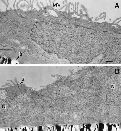
Figure 3.
Conventional electron microscopy of monolayers of cultured neonatal rat CP epithelial cells. Note abundant apical microvilli on the apical surface (A), the formation of basement membrane adjacent to the filter, and desmosomes between cells near their apical surfaces (B), features characteristic of native CP epithelia in vivo (18). MV, microvilli; B, basement membrane; N, nucleus; J, desmosome/tight junction complex. (Bar = 0.5 μm.)

