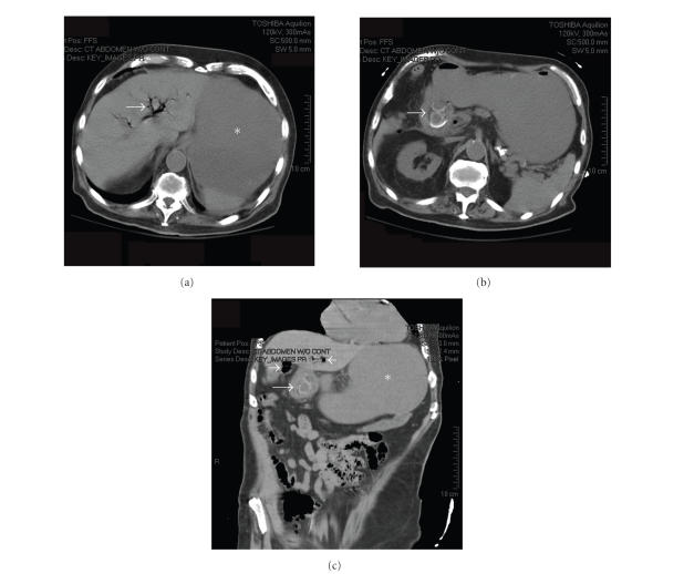
Figure 1.
Computed tomography (CT) scan of abdomen and pelvis. (a) Axial image showing pneumobilia (arrow) and a dilated fluid-filled stomach (). (b) 1-2 large gallstones (arrow) can be seen within an area of inflammation where the gallbladder is in close proximity to the duodenum. (c) Coronal reconstruction showing gallstone within duodenum (long arrow), Pneumobilia (short arrows), and dilated stomach () are also seen.

