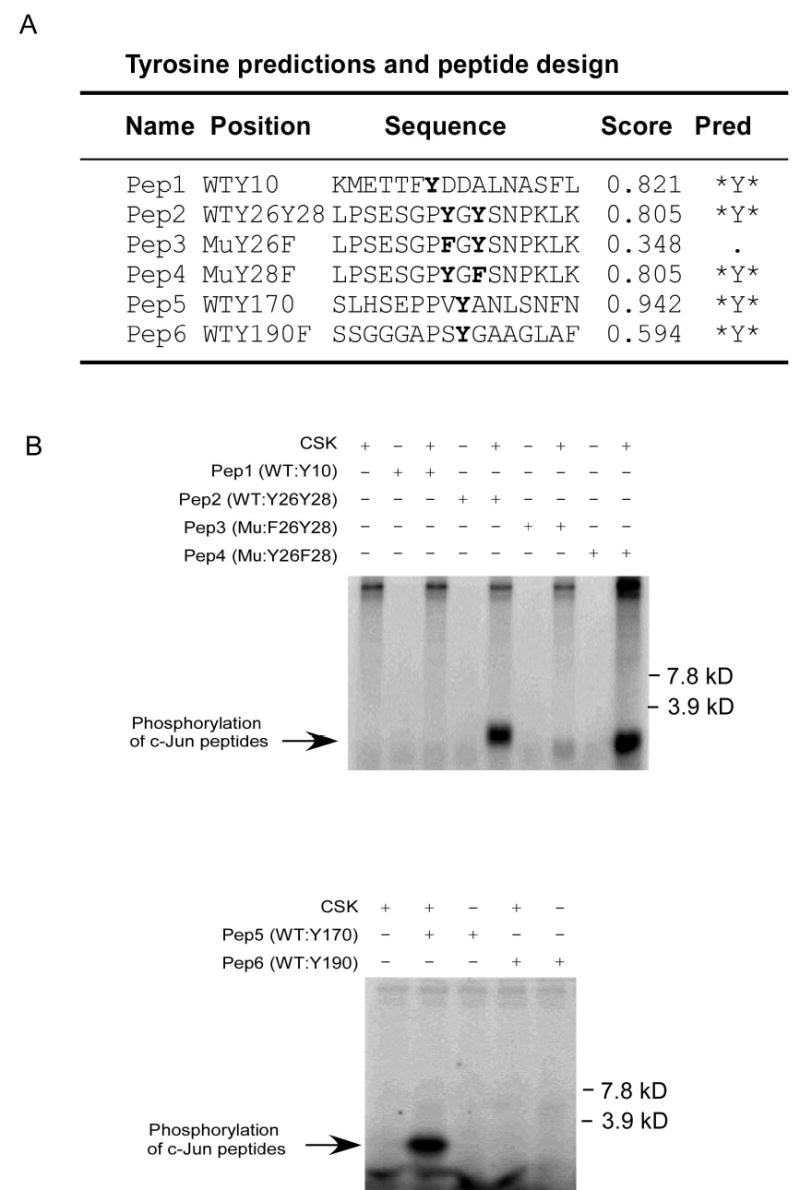
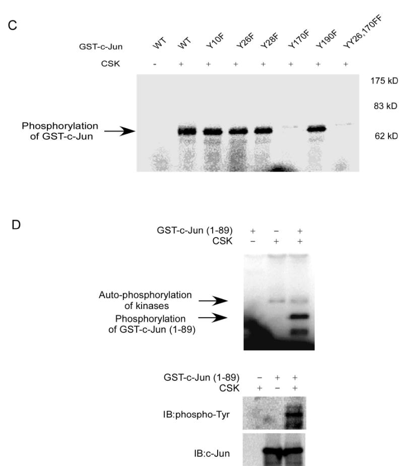
Figure 3.


Mapping of the c-Jun tyrosine site(s) phosphorylated by CSK. A, tyrosine sites predicted by NetPhos 2.0 and peptide design following prediction. WT: wild type; Mu: mutant; Score: possibility that the site might be phosphorylated in vivo; Pred: prediction. B, peptide mapping of the tyrosine sites phosphorylated by CSK as determined by an in vitro kinase assay in the presence of 32P as visualized by autoradiography. C, confirmation of the peptide mapping results by using GST-c-Jun or a GST-c-Jun-mutant as substrate in the in vitro kinase assay in the presence of 32P as visualized by autoradiography. D, confirmation of Y26 in c-Jun as a phosphorylation site of CSK using GST-c-Jun (1–89) as a substrate in an in vitro kinase assay in the presence of 32P as visualized by autoradiography or by Western blot.
