
Figure 4. Increased and delayed neurogenesis in adult THY-Tau22.
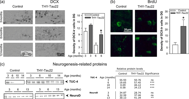
(a) DCX-ir in the DG of THY-Tau22 mice compared with WT at 3 months (n = 2), 6 months (n = 3 WT and n = 4 Thy-Tau22) and 12 months (n = 3). Representative images are shown. Note the age-related decay of DCX levels in control mice, while in THY-Tau22, increased numbers of DCX-ir cells were also observed at 6 months. Quantification of DCX-ir cells in the DG showed a significant increase at 6 months (*P < 0.05 at 6 months, n = 6 per genotype). Cell density is number of cells per field (photo taken with 10 × 10 magnification, approximately 190 × 100 μm). A coronal section (14 μm) showing the hippocampus around bregma −1.6 to −1.7 was used per animal. (b) 6-month-old mice were treated with BrdU i.p. for 7 days and sacrificed 14 days later. The number of BrdU-positive cells in the DG is significantly increased (*P < 0.05) in THY-Tau22 mice (n = 4) compared with littermate controls (n = 3). (c) Immunoblot analysis from hippocampal brain homogenates of proteins involved in neurogenesis and neuronal differentiation: TUC-4 is highly significantly and NeuroD mildly elevated in old THY-Tau22 mice compared with controls. Representative blots were shown and equal amounts loaded; similar results were obtained with n = 3–6 mice per genotype and band intensities quantified. Mean optical densities are shown in the table. Significance was calculated with two-way analysis of variance and Bonferroni’s post-test.