
Figure 2. Expression of EhPIG-M1 in E. histolytica WT and anti-sense strains.
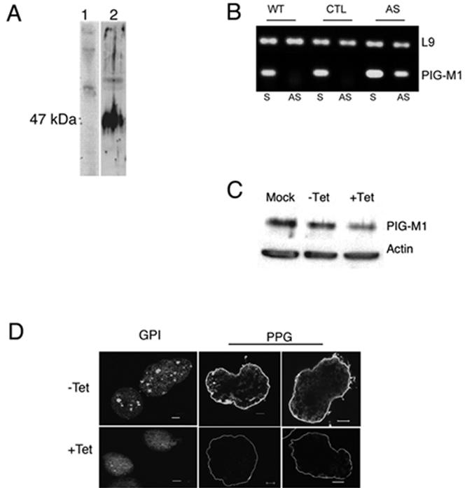
A. Protein analysis by immunoblot of extracts of wild type parasites. Electrophoretic resolution in 8% PAGE and immunoblots of proteins from a crude extract (equivalent to 104 cells by lane). Proteins were probed by immunoblot with pre-immune serum (lane 1) and with a anti-EhPIG-M1 antibody (lane2). B. Cloning and expression of anti-sense mRNA expressing parasites. The fragment cloned in anti-sense carries the first 708 bp of XM_64 49 88 loci. Agarose gel electrophoresis of PCR amplified products showed an unique DNA fragment (56 bp) amplified with antisense detecting primers when RNA was purified from PIG-M-AS strain. Antisense RNA was indeed absent from the wild type strain (WT) and from the control strain (CTL), which carries the vector. All strains allow for amplification of the sense mRNA. L9 correspond to ribosomal protein L9 encoding mRNA used as a control (81 bp). No amplification was detected for the two amplicons when RT was absent in the assay. S = sense detecting primers, AS = anti-sense detecting primers. C. Western blot analysis of protein lysates separated in SDS-PAGE. Extract from PIG-M-AS parasites (+/− tetracycline) were used (equivalent to 104 cells by lane). EhPIG-M1 is at 47 kDa, and actin used as a blot reference at 43 kDa. The ECL film image was treated with ImageQuant software. D. Labeling of GPI-linked molecules by FLAER and PPG by immunofluorescence. PIG-M-AS cells grow with or without Tet were incubated with FLAER and treated for confocal microscopy. Upper left panel: FLAER labeled uninduced cells. Lower left panel: FLAER labeled induced cells expressing antisense mRNA, notice the loss of vesicle labeling. To localize PPG , PIG-M-AS trophozoites growing in the presence (or not) of tet for 5 days were fixed and incubated with specific mAb-5 recognizing PPG and treated for confocal microscopy. The micrograph (taken at the middle of the cell) shows a reduction of PPG labeling when the AS-cells were treated with tet. Notice the lost of spike-like structures around the cell. Bar = 5 m.