
Figure 3. GPI-intermediate biosynthesis with extracts from EhPIG-M1 antisense blocked cells.
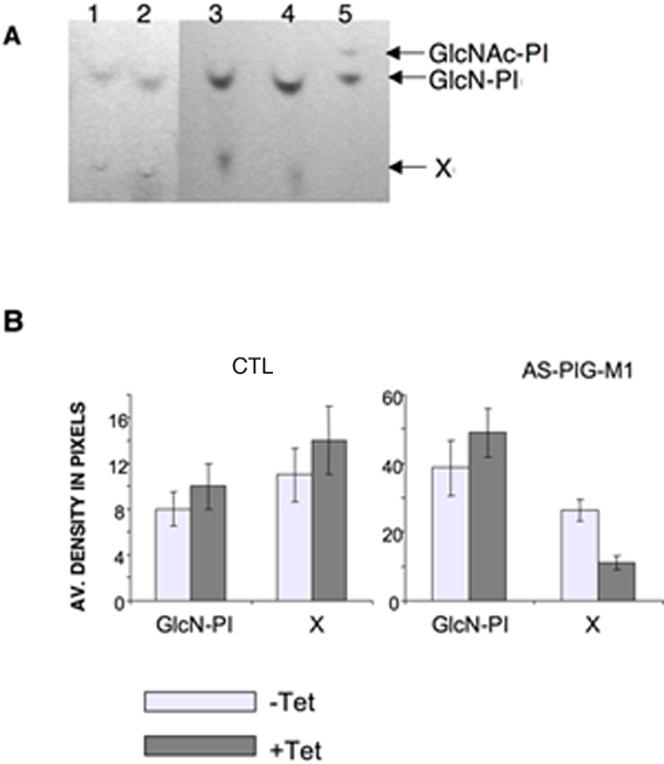
Crude membrane preparation from tetracycline induced and uninduced trophozoites were labeled with [14]-C UDP-N-acetylglucosamine in a cell free system in the presence of GDP-mannose. GPI biosynthesis intermediates were isolated and analyzed by HPTLC and visualized by fluorography. Panel A, Lane 1-2: GPIs from untreated and tetracycline treated vector control respectively, lane 3-4, GPIs from untreated and tetracycline treated EhPIG-M1 antisense cells line respectively; lane 5 shows the first two intermediates of the GPI-pathway, namely GlcNAc-PI and GlcN-PI, as control. Panel B shows the densitometric analysis of the radiolabeled products as obtained on HPTLC chromatography, I- GlcN-PI and product X for untreated (clear boxes) and tetracycline treated (black boxes) cells carrying the control vector (CTL), or the EhPIG-M1 antisense construct. An increased level of GlcN-PI (the substrate of PIG-M) is observed in extract carrying reduced levels of EhPIG-M1.