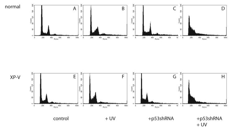
Figure 2.
Flow cytometric analysis of normal (GM5659DT) and XPV (GM3617T) cells irradiated with 0 (A,B,C,D) or 5.2 J/m2 of UV (C,D,G,H) and harvested 16 hours later and stained with propidium iodide. G1 and G2 peaks identified in upper left panel. A: GM5659DT cells infected with pUper.Retro.Neo.Gfp vector. E: unirradiated GM3617T cells infected with pSuper.Retro.Neo.Gfp vector. B: UV-irradiated GM5659DT cells infected with pSuper.Retro.Neo.Gfp vector. F: GM3617T cells infected with pSuper.Retro.Neo.Gfp vector, grown in medium after UV. C: UV-irradiated GM5659DT cells infected with pSuper.Retro.p53.Neo.Gfp or lower, unrradiated pSuper.Retro.p53.Neo.Gfp GM3617T cells. D: UV-irradiated GM5659DT cells infected with pSuper.Retro.p53.Neo.Gfp or H, UV-irradiated pSuper.Retro.p53.Neo.Gfp GM3617T cells.

