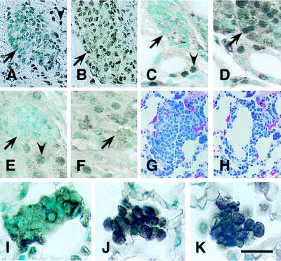
Figure 2.
Characterization of RB transgene expression. (A–F) Tetracycline-regulated expression of RB in Rb+/−RTgRB mice. Transgenic littermates were treated with tetracycline hydrochloride (1 mg/ml) from fertilization until either P94 (A, C, and E) or P90 (B, D, and F). On P94 expression of the RB transgene was tested in atypical cells of the pituitary intermediate lobe (A and B), the thyroid gland (C and D), and the adrenal medulla (E and F). In mice continuously exposed to tetracycline no RB is detected in nuclei of atypical (arrows) melanotrophs (A), C cells (C), and medulla cells (E). Withdrawal of tetracycline on P90 results in expression of RB transgene in those cells by P94 (B, D, and F). Note that staining of normal cells reflects expression of either endogenous Rb (A, C, and E, arrowheads) or both endogenous Rb and exogenous RB (B, D, and F). (G–K) RB expression in metastatic C cells after administration of LPDs. Mice were killed 24 hr after i.v. injection with either LPD-RB (G, H, and J), LPD-RBH209 (K), or LPD-lacZ (I). (G and H) Groups of metastatic cells before (G) and after (H) microdissection. (I–K) Simultaneous detection of cytoplasmic calcitonin and nuclear RB in metastatic cells. Unlike metastatic cells from mice treated with LPD-lacZ (I), those from mice treated with either LPD-RB (J) or LPD-RBH209 (K) showed intense RB staining. Avidin-biotin-peroxidase immunostaining with C-15 antiserum alone (A–F) or together with calcitonin-specific antibodies (I–K). Counterstaining with methyl green. (G and H) Hematoxylin-eosin staining. [Bar: 40 μm (A and B), 20 μm (C–F), 55 μm (G and H), and 16 μm (I–K).]

