Figure 5.
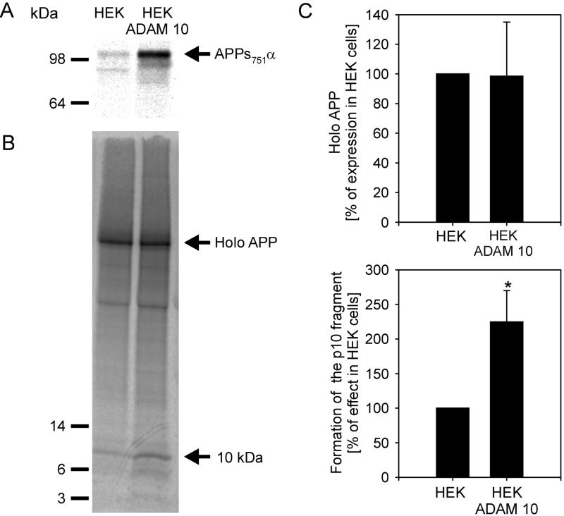
Effect of ADAM 10 on the production of APPsα and on the 10-kDa C-terminal fragment. (A) Cells were metabolically labeled with l-[35S]methionine and [35S]cysteine (200 μCi/ml) for 5 hr. Cell media were immunoprecipitated with antibody 1736 and analyzed by SDS/PAGE in 10% gels. (B) Cell lysates were immunoprecipitated with antibody C7. The samples were separated by 10–20% Tris/Tricine gel (Novex) and analyzed as described. (C) Quantitative analysis of holo-APP and p10. The values of p10 were normalized to the levels of holo-APP. The results are the averages ±SD of four experiments. Statistical significance between control cells and HEK ADAM 10 cells was determined by Student’s unpaired t test (∗, P < 0.005).

