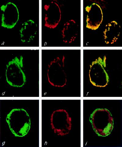
Figure 3.
Colocalization of HA-PKD2 and TRPC1 in live cells. HEK293T cells were cotransfected with HA-PKD2 and F-TRPC1 (a–c), HA-PKD2 and M-TRPC1 (d–f), or HA-PKD2 and TRPC3-M (g–i), and the subcellular distribution of the encoded proteins was determined by double immunofluorescence using confocal microscopy. FLAG- or myc-tagged proteins were stained with a FITC-conjugated secondary antibody, while HA-PKD2 was stained with rhodamine-conjugated secondary antibody. Computerized images of green fluorescein staining corresponding to F-TRPC1, M-TRPC1, or TRPC3-M are shown in a, d, and g, respectively, whereas red rhodamine images corresponding to HA-PKD2 are shown in b, e, and h. Fluorescein and rhodamine merged images corresponding to HA-PKD2 and F-TRPC1, HA-PKD2 and M-TRPC1, or HA-PKD2 and TRPC3-M subcellular distributions are shown in c, f, and i, respectively.

