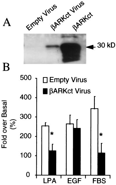
Figure 1.
Effect of βARKct on MAP kinase activation in rat aorta VSM cells. (A) Representative Western blot demonstrating that the βARKct peptide is expressed in cellular extracts only after βARKct adenovirus infection. Shown in the right-hand lane of this blot is a βARKct-positive standard that is a COS cell extract overexpressing the ∼30-kDa βARKct peptide. (B) Histograms displaying MAP kinase activity expressed as the fold over basal activity in rat aorta VSM cells infected with either the βARKct adenovirus or an empty virus as a negative control. MAP kinase activity was induced by LPA (10−5 M), EGF (10−5 M), or serum (5% FBS). Data shown are the means ± SEM of five separate experiments done in triplicate. ∗, P < 0.05 βARKct versus empty virus (paired t test).

