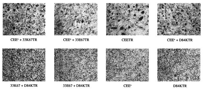
Figure 6.
Chimeric envelope protein-mediated syncytia formation of 3T3/CD33 cells. Envelope protein expression plasmids were cotransfected (at a ratio of 1:1) into 3T3/CD33 cells. At 36 hr after transfection, the cells were stained with methylene blue, and those cells containing more than four nuclei counted as syncytia. (Upper) Examples of positive syncytia formation; (Lower) Examples of negative syncytia formation. These data are a portion of those summarized in Table 2.

