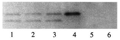
Figure 7.
Heterooligomer formation between chimeric envelope monomers and wild-type monomers. SDS/PAGE (14%) of the coimmunoprecipitated virion envelope protein is shown. Envelope proteins were transiently expressed in 293T cells. Metabolic labeling for 4 hr with [35S]Met was performed 24 hr after transfection. The cells were lysed, and the supernatant was immunoprecipitated with 5 μ of anti-R peptide (22). The p12E protein, bottom band, that was present in 33E67TR and 33K67TR, was immunoprecipitated by R-peptide antiserum only in the presence of coexpressed D84K (lane 1, 33E67TR/D84K; lane 2, 33K67TR/D84K); or CEE+ (lane 3, 33K67TR/CEE+), but not when expressed by itself (lane 5, 33E67TR and lane 6, 33K67TR). There is no p12E band in the wild-type CEE+ (lane 4), indicating that there is no R-peptide cleavage in this 293T system.

