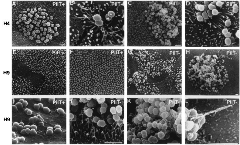
Figure 2.
SEM examination of a T84 monolayer infected for 4 h (A–D) or 9 h (E–L) by PilT+ (A, B, E, F, and I) or PilT− bacteria (C, D, G, H, J, K, and L). Initially, both strains have a similar pattern of adhesion (A–D); however, after a longer incubation period, PilT+ spread onto the monolayer (E and F) and adhere very intimately to cells (I). In contrast, PilT− bacteria remain as large, piliated clumps sitting on the cells (G, H, J, K, and L) (Bars = 2 μm.)

