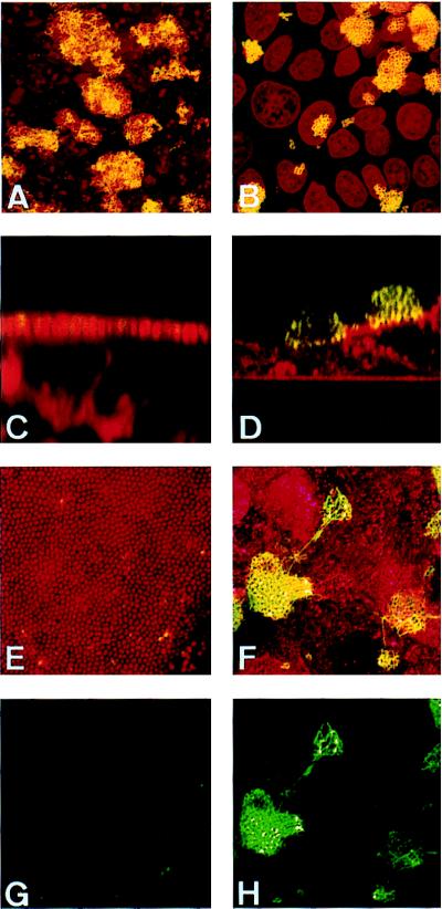
Figure 3.
Confocal immunofluorescence micrographs of T84 cells grown on coverslips infected for 4 (A and B) or 9 (C–H) h by clone12pilT+ (A, C, E, and G) or by clone12pilT− (B, D, F, and H). MC and eukaryotic cells were stained with ethidium bromide (A, B, C, E, and G) or with tetramethylrhodamine B isothiocyanate-labeled lectin from Triticum vulgaris (D, F, and H) (shown in red). Pili were labeled using the 20D9 mAb (shown in green). A–F corresponds to the superimposition of both stainings. G and H display only pili staining of adherent MC. A, B, E, F, G, and H are images reconstructed from confocal xy sections of infected monolayers, and C and D are from xz sections.

