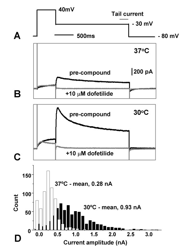
Figure 2.
Increased functional expression of hERG at 30°C. HERG-expressing CHO cells were grown at 37°C (Materials and Methods), and then split and kept subconfluent at either 37 or 30°C for 3 d. Voltage command protocol (A). HERG currents were averaged from the last 200 mS of the -30 mV inactivation step before compound addition, subtracting the same recording after the application of 10 μM dofetilide. Patch clamp recordings made with an IonWorksHT instrument from a representative cell maintained at either 37 or 30°C (B,C, respectively). The black traces are recordings before compound addition. The grey traces are recordings following 10 μM dofetilide addition. It is noticeable that the difference at the initial phase of the -30 mV step was even bigger between the two temperatures. Population analysis of the hERG currents using three 384 well patch plates each (ie. >1000 recordings) for cells grown at 37 (open bars) and 30°C (black bars; D).

