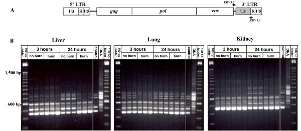
Figure 1.
Changes in mRNA expression profiles of MuERVs in distant organs after burn. (A) Schematic representation of primer locations on MuERV. A set of primers (ERV-U2 and ERV-U1) flanking the 3' U3 region are indicated by arrows. (B) RT-PCR analysis of the MuERV expression after burn in the liver, lung, and kidney. Tissues (liver, lung, and kidney) harvested at 3 hours and 24 hours after 18 % TBSA burn were subjected to RT-PCR analysis of MuERV expression using a primer set flanking the 3' U3 region. Respective tissues harvested without any treatment, except for cervical dislocation, serve as a no treatment control in comparison to no burn controls (subjected to anesthesia, resuscitation, and CO2 inhalation). One representative sample out of three no treatment controls for each tissue is presented. In addition, a genomic MuERV profile was used as a reference.

