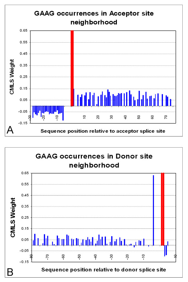
Figure 7.
The weight distribution of the ESE motif GAAG in the acceptor (A) and donor (B) splice-site neighborhood. The x-axis shows the acceptor splice-site neighborhood interval. The consensus dinucleotide AG location is marked with the red bars (positions -1 and -2) in Figure 7A. The consensus dinucleotide GT location is marked with the red bars (positions 1 and 2), in Figure 7B. For every occurrence of the feature GAAG in the set A-4mer [-80,80], we draw a bar corresponding in height to its CMLS assigned weight. This feature has a negative weight when it is positioned in the intron region, but a positive weight downstream the splice site. For the donor site, we notice its exceptionally high weight at position -4. One possible reason may be the reflection of the donor-site consensus signal.

