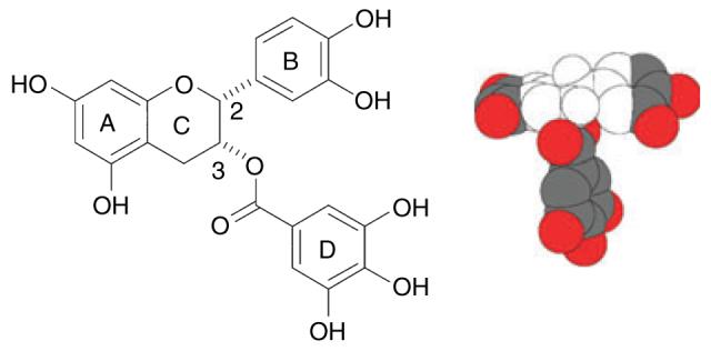
Figure 1.

Structure of (−)-epicatechin gallate (ECg), showing the B- and D-ring pharmacophores. The hydrophobic and hydrophilic domains of ECg are indicated in the space-filling model (provided by J.C. Anderson, University of Nottingham, UK) by the colourless and coloured regions, respectively.
