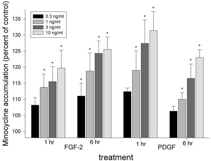
Figure 1.
Stimulation of fibroblast minocycline accumulation by FGF-2 and PDGF. Confluent fibroblast cultures were starved for 20 hrs and treated with the indicated growth factor concentrations for 1 or 6 hrs. Cell DNA content did not increase significantly under these experimental conditions. After brief incubation at 37°C, 40 μg/mL minocycline was added, and uptake was monitored for 3 min. The data represent the mean ± SEM of 5 individual experiments. Both agents produced a significant treatment effect after 1 and 6 hrs (P < 0.003, repeated-measures ANOVA). Conditions that produced a significant increase in minocycline accumulation compared with controls (P > 0.05, Dunnett’s test) are indicated by *.

