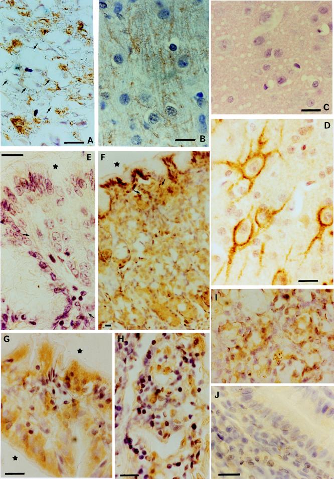
Figure 1.
(A) Zoo lemur no. 703. PrP deposits in large vacuolated fibers of the ventral funiculus of the cervical spinal cord. Arrows point to fiber membranes. Anti-PrP 3F4, 1:200. (B) Zoo lemur no. 712. Nerve fibers showing PrP immunoreactivity (brown) in layer IV of the cerebral cortex. Anti-PrP 3F4, 1:200. (C) Experimental BSE-infected microcebe no. 656. Microvacuolation in the neuropil of the parietal cortex (layer V). Hematoxylin and eosin. (D) Experimental BSE-infected microcebe no. 656. Abnormal Tau proteins inside pyramidal neurons of the parietal cortex layer III. Anti-tau 961S28T, 1:200. (E) Experimental control microcebe no. 593. High magnification of the stomach wall: no PrP immunoreactivity is detected in the epithelium, secretory glands, or various lymphoreticular tissue elements (arrows). Star indicates luminal surface. Anti-PrP 3F4, 1:200. (F) Experimental BSE-infected microcebe no. 656. PrP distribution in the stomach wall. Arrows point to reticulolymphatic elements; star indicates luminal surface. Anti-PrP 3F4, 1:500. (G) Experimental BSE-infected microcebe no. 656. PrP localization in an intestinal villus. Note the interrupted epithelium at the level of M cells containing a lymphocyte, and the immunoreactivity of lymphoid reticular structures. Stars indicate luminal surfaces. Anti-PrP106–126, 1:2. (H) Experimental BSE-infected microcebe no. 656. Peyer’s patch with PrP immunoreactive lymphoid structures. Anti-PrP106–126, 1:2. (I) Experimental BSE-infected microcebe no. 656. PrP labeling in splenic red pulp. Anti-PrP 3F4, 1:500. (J) Experimental BSE-infected microcebe no. 656. Small intestine. Anti-PrP 3F4, 1:200, pre-adsorbed with PrP antigen.

