
Figure 1. Expression of ANGPTL4 in mice ischemic hindlimbs.
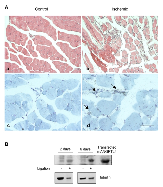
Ischemic and controlateral tibialis anterialis muscles were harvested at day 2 or 6 post ligation of the left femoral artery. A, At day 6, non ischemic muscle shows normal histology with Hematoxylin and Eosin staining (a) and no detectable signal in vessels nor muscle fibers after in situ hybridization with the FIAF antisense probe (c). Ischemic muscle shows fiber necrosis, interstitial oedema and inflammatory infiltrate (b). Signal is detected in small vessels (arrows) in ischemic areas with the FIAF antisense probe (d). Bar = 100 μm. B, Western-blot analysis of muscle protein extracts (100 μg/lane) with anti-mouse ANGPTL4 (top panel) or anti-tubulin (bottom panel). Transfected mouse ANGPTL4 was used as a positive control.