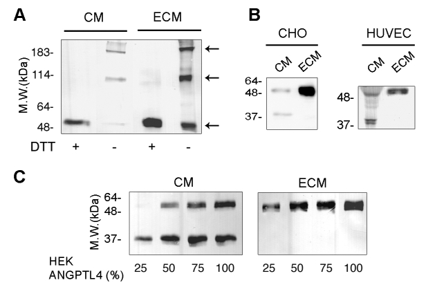
Figure 3. Accumulation of full-length ANGPTL4 in the ECM.

A, CM or ECM from CHO-DHFR-ANGPTL4-myc cells was analyzed by Western-blot using anti-myc antibody on 7.5% SDS-PAGE in reducing (+DTT) or non-reducing (−DTT) conditions. B, CM or ECM from CHO-DHFR-ANGPTL4-myc cells or transfected HUVEC was analyzed by Western-blot using anti-myc antibody. C, Increasing ratios of HEK293-ANGPTL4-flag versus HEK293 cells were plated in the indicated proportions and grown until confluence. CM (left panel) and ECM (right panel) were analyzed by Western-blot using anti-flag antibody. Results are representative of three independent experiments.
