FIGURE 4.

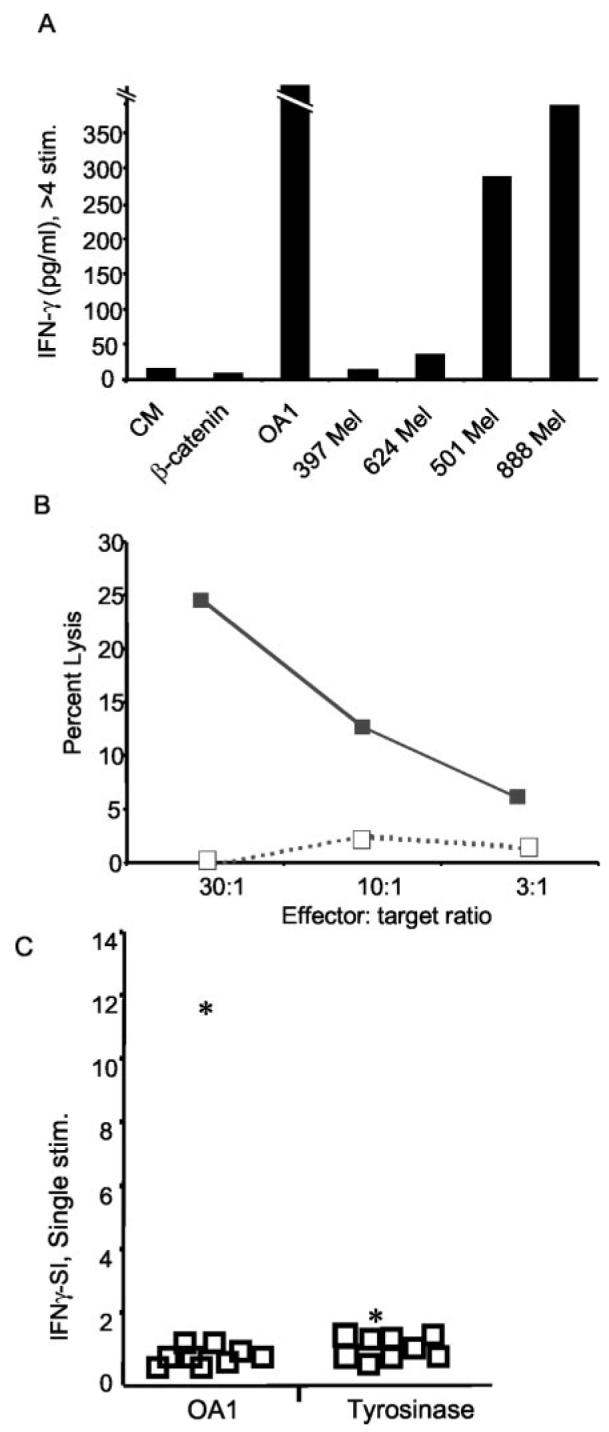
T cells from the OA1 Ag-loss variant patient avidly respond to OA1 and recognize melanoma. A, Long-term CD8+ T cells from line SE OA1-3 2/6, generated by four or more in vitro stimulations with OA1126–134, recognize OA1 peptide and A*2402+/OA1+ melanoma lines (501 and 888 Mel), but not A*2402−/OA1+ melanoma lines (397 and 624 Mel), nor the control peptide (mutated β-catenin29–37) pulsed onto 888 EBV-B. B, T cell line obtained by repeated stimulation of PBMC from patient OAP-46 with peptide OA1126–134 lyse A*2402+/OA1+ melanoma 888 (■), but not A*2402−/OA1+ melanoma 624 mel (□) in a 4-hour 51Cr-release assay. C, T cells from patient OAP-46 have superior reactivity to OA1. T cells from patient OAP-46 react with OA1126–134, but not tyrosinase 205–213, while T cells from normal volunteers and melanoma patients lack reactivity to both Ags. PBMC from nine normal volunteers (□) and OA1 patient OAP-46 (*) were stimulated for 7 days in vitro with either OA1126–134 or tyrosinase 205–213. To evaluate responses from all samples to both Ags, T cells were stimulated with OKT3 (anti-CD3), control peptide (mutated β-catenin 29–37 pulsed at 10 μM), or the specific peptide in question (either OA1126–134 or tyrosinase 205–213 also pulsed at 10 μM) both pulsed onto 888 EBV-B. All lines secreted IFN-γ in response to OKT3 (not shown). The IFN-γ-SI was generated by dividing the IFN-γ secretion produced to OA1126–134 or tyrosinase 205–213 against the secretion produced against mutated β-catenin 29–37. Experiments in all panels were performed three times with similar results, using different but comparable targets to demonstrate the generalizability of the findings. CM, culture media.
