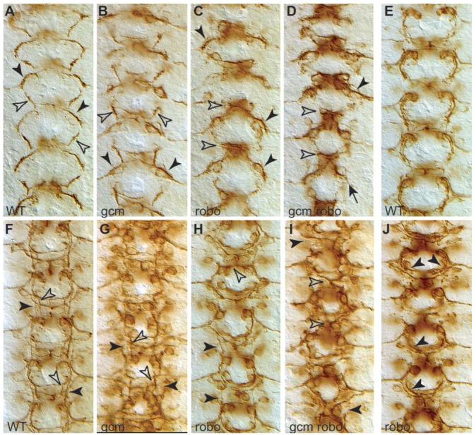
Fig. 5.

Axonal defects occur from the earlier phases of pioneer axon extension. Pioneer axons visualised with 22c10 antibodies in wild-type (A,E,F), gcm-mutant embryo (B,G), robo1 mutant (C,H,J) and gcm-robo1 double mutant (D,I) at stages 12.1 (A,B,C,D); 13 (E,J) and 14 (F,G,H,I). (A) In wild-type embryos, the descending dMP2/MP1 axons (black arrowhead) meet the ascending vMP2/pCC axons (white arrowhead) at the end of stage 12. (B) In gcm mutants, the dMP2/MP1 fascicle misroutes to the muscle, so its descending trajectory is missing (black arrowheads), but the vMP2/pCC fascicle is unaffected (white arrowheads). (C) In robo mutants, the pCC/vMP2 fascicle does not grow but is trapped in the midline (white arrowheads), whereas the dMP2/MP1 fascicle begins its normal descending trajectory. This is also seen slightly later, at stage 13, (J) where the dMP2/MP1 fascicle extends normally all the way down (arrowheads) and only then curves towards the midline as it fails to encounter the ascending vMP2/pCC fascicle, which is missing (compare to E, wild-type). (D) In gcm-robo1 double-mutant embryos, the pCC/vMP2 fascicle is completely collapsed on the midline (white arrowheads) and the dMP2/vMP2 fascicle is either missing or misrouted to the muscle (black arrowheads). (F) At stage 14, the two fascicles, dMP2/MP1 (black arrowheads) and pCC/vMP2 (white arrowheads) are clearly distinguishable. (G) In gcm mutants, only the pCC/vMP2 fascicle is present (white arrowheads) and the dMP2/MP1 fascicle is missing (black arrowheads). (H) In robo1 mutants, the pCC/vMP2 fascicle collapses on the midline (white arrowhead) but the dMP2/MP1 fascicle (black arrowhead) is normal. (I) In gcm-robo1 double mutants, the pCC/vMP2 fascicle collapses on the midline, but the dMP2/MP1 fascicle is not discernible. Anterior is up.
