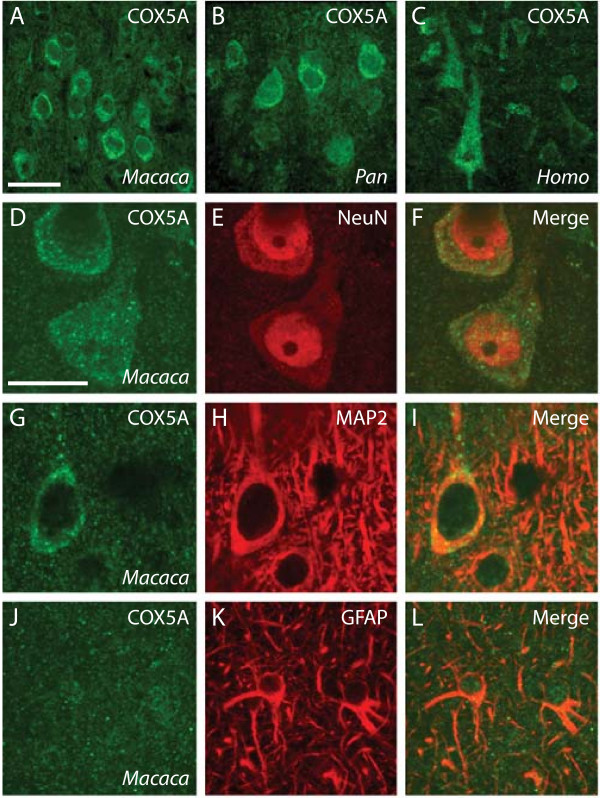
Figure 4.
Immunohistochemical staining of COX5A protein in the dorsolateral prefrontal cortex. The distribution of staining using a monoclonal antibody against COX5Ap in macaque monkey (A), chimpanzee (B), and human (C). Panels D-L show double label immunostaining from macaque monkey. Images in A-I are taken from layer III. Images in J-L are taken from layer I. Scale bar in panel A = 50 μm applies to A-C. Scale bar in D = 25 μm applies to D-L.

