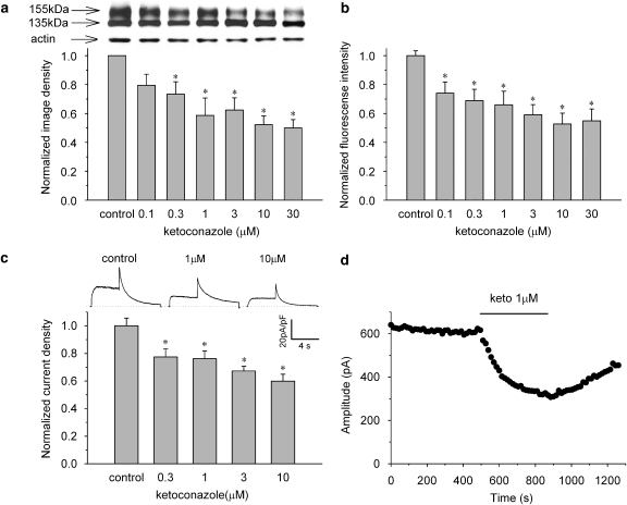
Figure 3.
Ketoconazole reduced maturation and surface expression of hERG channels. Concentration-dependent decrease of 155-kDa hERG protein (a), fluorescence intensity of antibody-labelled hERG channels detected by flow cytometry (b) and current densities (c). The cells were incubated for 48 h in control or ketoconazole-containing medium. Panel a, upper panel: representative western blot analysis. Data are mean±s.e.m. *P<0.05, compared with the control (n=6). Panel b, data are mean±s.e.m. *P<0.05, compared with the control (n=9). Panel c, upper lane: representative current traces under control conditions and after long-term application of ketoconazole at 1 and 10 μM (see Figure 2a for pulse protocol). The hERG current density was measured after 1 h following the washing of the drug. Peak tail current densities at each concentration were normalized to the value in control conditions. Data are mean±s.e.m. *P<0.05, compared with the control (n=16–21). (d) Time course of direct hERG current block by 1 μM ketoconazole after 48-h incubation with 1 μM ketoconazole. Amplitude of the peak tail current was plotted against time (see Figure 2a for pulse protocol). An acute hERG current block occurred after disruption of hERG protein trafficking.

