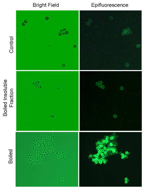
Figure 6.
Epifluorescent images of polyclonal anti-Cryptosporidiu m antibody binding to oocyst walls. Oocysts were labeled with rabbit polyclonal antibody, which was then tagged with FITC-labeled anti-rabbit IgG. Oocysts were viewed and imaged using bright field and an Olympus UM-WIBA FITC fluorescence filter (400× magnification).

