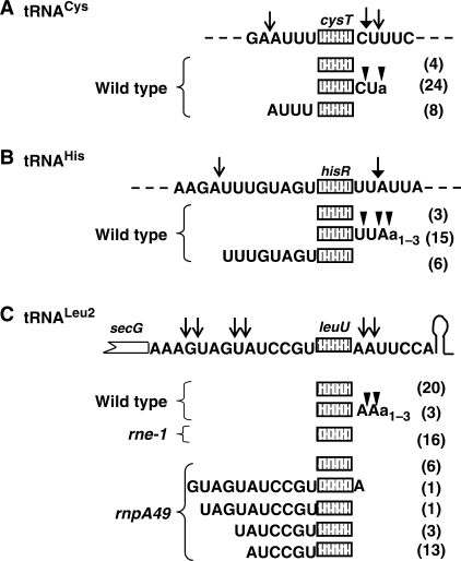
Figure 5.
Identification of 5′ and 3′ ends of tRNACys (A), tRNAHis (B) and tRNALeu2 (C) using RT-PCR cloning of 5′–3′ end-ligated transcripts. The total RNA was self-ligated, reverse transcribed, amplified using cysT, hisR and leuU-specific primers, cloned and sequenced as described in the Materials and Methods section. A schematic representation of each tRNA with up and downstream sequences is shown directly above respective tRNA clones. RNase E cleavage sites as identified previously (solid arrow) and in this study (open arrow) are indicated at the top of each diagram. Multiple 3′ ends (inverted triangles) with similar 5′ ends were obtained for various tRNA clones. Numbers in the parenthesis to the right of each diagram are the number of clones sequenced for that species. Lower case a's indicate the presence of non-templated residues.

