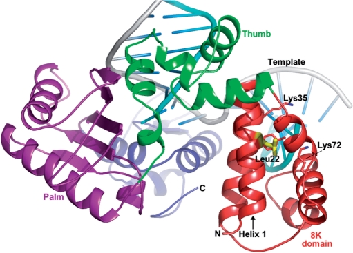
Figure 1.
A ribbon representation of polymerase beta. The structure of pol β is depicted in domain colors showing the dRP lyase domain (8 kDa domain) in red, the N-terminal/thumb domain in green, the palm domain in magenta, the C-terminal/fingers domain in blue, the DNA template in gray and the downstream and primer strands in cyan. Leucine 22, shown in yellow, is located ∼11 Å away from the dRP lyase catalytic site (Lys72). Critical dRP lyase active site residues (Lys72, Lys35 and Tyr39) are shown in atom colors (carbon, red; nitrogen, blue).

