
Figure 7.
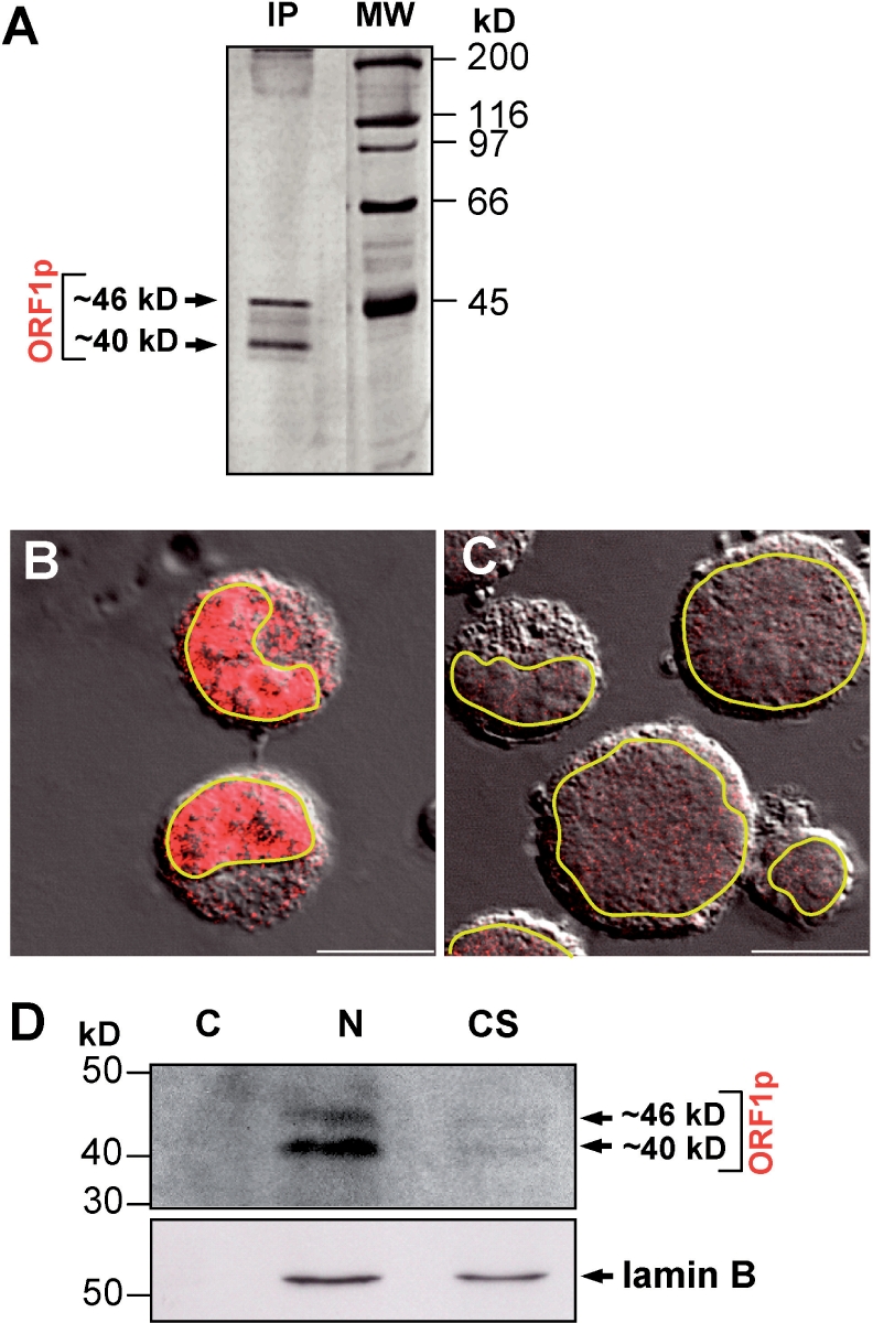
Endogenous L1Rn ORF1-encoded proteins are predominantly localized to the nucleus of RCL cells. (A) Silver staining of L1Rn ORF1-encoded proteins immunoprecipitated from RCL cell lysates with RG24 IgG. Molecular weights of the two proteins of ∼46 and ∼40 kDa correspond to the theoretical masses of L1Rn-ORF1 protein classes I-21a and I-21p, respectively. (B) Confocal images of RCL cells immunostained with the rG24 antibody (red). (C) Secondary antibody control. Confocal images were merged with a differential interference contrast (DIC) micrograph to show the outlines and nuclei of cells. Outlines of nuclear envelopes are emphasized in yellow. Scale bar—10 µm. (D) Immunoblot analysis of subcellular fractions of RCL cells. Three different protein fractions (C, cytosolic; N, nuclear; CS, cytoskeletal matrix proteins) were separated by SDS–PAGE and blotted onto nitrocellulose. The membrane was incubated consecutively with anti-ORF1p- antibody (upper panel) and with an antibody directed against the nucleus-specific laminB protein (lower panel), which shows efficient separation of the subcellular fractions.