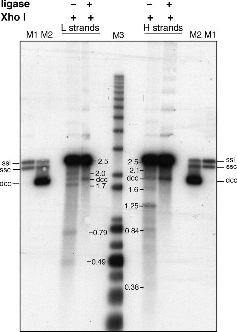
Figure 4.
Ligation of discontinuities in kinetoplast-associated minicircles. Purified kDNA from 300 min after release from the hydroxyurea block were either treated with E. coli DNA ligase (+) or not (−) prior to digestion with XhoI and alkaline denaturing gel analysis. The gel was transferred and hybridized as in Figure 3 to identify released H and L single strands. Marker lanes: M1, minicircle single strand linears (ssl) and single strand circles (ssc); M2, denatured covalently closed minicircles (dcc) in addition to minicircle single strand linears (ssl) and single strand circles (ssc); M3, molecular weight markers.

