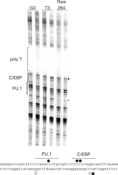
Figure 5.
In vivo DMS footprinting of the csf1r promoter using PAP-LMPCR. Radiolabelled PAP-LMPCR products from genomic DNA purified from DMS-treated NIH3T3 fibroblast cells (csf1r non-expressing) or RAW264 macrophage cells (csf1r expressing) as well as DMS-treated genomic DNA as indicated were run on a 6% sequencing gel. The experiment shows clear footprints within the target region in RAW264 cells closely associated with the C/EBP- and PU.1-binding sites. Samples are in duplicates, demonstrating the reproducibility of the reaction. Footprints are marked with either closed circles for guanines showing DMS hyper-reactivity or open circles indicating hypo-reactivity. The lower panel shows the position of footprints previously demonstrated on the upper strand (18); the footprints on the lower strand are from the experiment described here.

