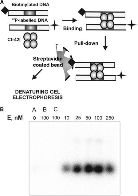
Figure 4.
DNA synapsis by Cfr42I. (A) Schematic representation of the biotin pull-down assay. The reactions contained equimolar amounts of two specific 30 bp duplexes (10 nM each) with various amounts of Cfr42I. The first of the duplexes carried a biotin tag and the other was radiolabelled with 32P. After 5 min preincubation of enzyme and DNA, streptavidin-coated magnetic beads were added that adsorbed the biotin-tagged DNA. The beads were harvested and the amount of radiolabelled DNA associated with the beads was measured after denaturing PAGE. The radiolabelled DNA is pulled down with the beads only if Cfr42I forms complexes with two DNA molecules. (B) Results of the pull-down assay. Cfr42I concentrations were as indicated above each lane. The gel lanes marked ‘A’, ‘B’ and ‘C’ are control pull-down experiments performed in the absence of protein (lane ‘A’), in the presence of non-specific biotinylated DNA (lane ‘B’) or in the absence of biotinylated DNA (lane ‘C’).

