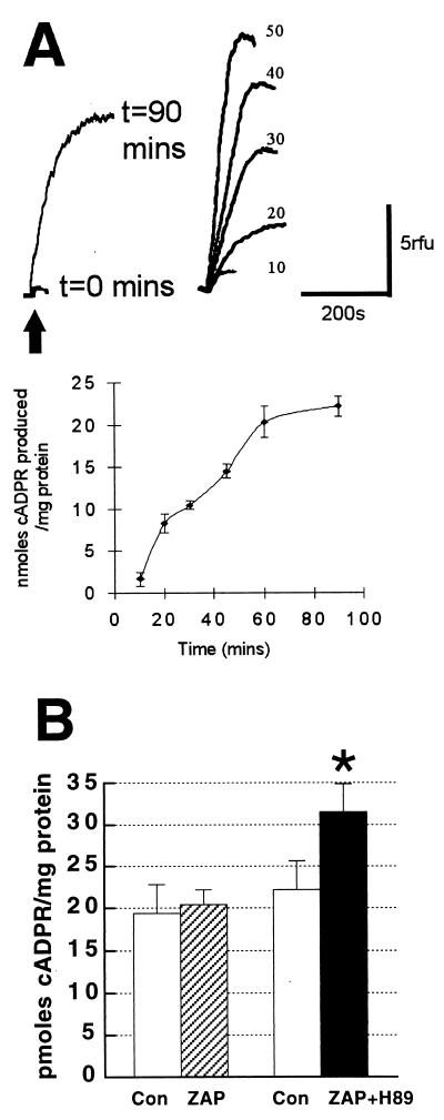
Figure 2.
Hippocampal slices possess cGMP-stimulated ADP-ribosyl cyclase activity. (A) (Upper) Fluo-3 fluorescence (relative fluorescence units; rfu) increases in sea urchin egg homogenate produced by known concentrations of cADPR (arrow). Numbers next to each trace are cADPR in nmol, against which hippocampal cADPR activity was calibrated. (Lower) Time course of cADPR synthesis in hippocampal homogenates incubated with 2.5 mM β-NAD+ at 37°C. After the indicated times, reaction was stopped and 5 μl of incubation medium was added to sea urchin homogenate (n = 6) for fluorescence measurement of amount of cADPR. (B) Mean ± SEM amount of cADPR in control hippocampal slices (open bars), versus slices treated with 20 μM zaprinast (ZAP; hatched bar), a type V phosphodiesterase inhibitor that raises cGMP concentration by preventing its degradation, and 20 μM zaprinast plus the PKA inhibitor H89 (10 μM; solid bar). n = 12–14 slices per group in three experiments. ∗, P < 0.05, Student’s t test compared with control slices.

