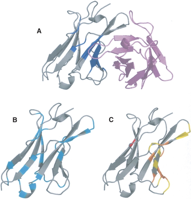
Figure 5.
Jto, a variable domain from λ-6 type immunoglobulin light chain with 111 residues (1CD0). (A) Seventeen HFV residues are colored blue, displayed in complex. (B) The conserved residues are in cyan. (C) The main binding site is colored yellow, the surface patches composed of nine residues in two patches with eight and one residues are colored red, and the intersection is colored orange.

