
Figure 6.
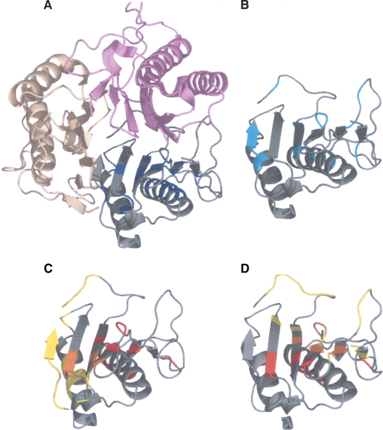
Yjgf protein (1QU9) with 126 residues. Twenty-one HFV residues are determined, 19 are grouped into one cluster. The surface patches determined are composed of 12 residues clustered into groups of 10 and 12. (A) The HFV residues are colored blue; the complex partners are in two different shades of pink. (B) The conserved residues are in cyan. (C) The main binding site is in yellow, the surface patches are colored red, and the intersection colored orange. (D) The second binding site is colored yellow, the surface patches colored red, and the intersection is colored orange.