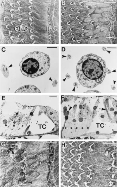
Figure 3.
Morphological comparison of the organ of Corti of adult p27+/+ (wild-type) and p27−/− (homozygote) mice. SEMs of the apical region of p27+/+ (A) and p27−/− (B) are shown. Note that p27−/− has the fairly typical pattern of three to four rows of OHCs and one row of IHCs as seen in the p27+/+. However, additional (arrow) and abnormal-appearing hair-cells (arrowhead) are seen in p27−/−. Also note that in the region between the IHCs and first row of OHCs in p27−/− there is disorganization of the normal pattern of rectangular apices of the inner pillar cells, with a higher density of irregular apices. (C) In p27+/+ at the level of the OHC nuclei, two Deiters’-cell phalangeal processes (arrows) normally can be seen adjacent to a single OHC. (D) In the p27−/− mice a single OHC is surrounded by up to six Deiters’-cell phalangeal processes (arrows). (E) Radial sections of p27+/+ show normal pillar-cell and phalangeal-cell morphology and number, with three OHC rows and three Deiters’-cell rows (∗), with normal spaces of Nuel and tunnel of Corti (TC). (F) In radial sections of p27−/−, there is an increase in the number of Deiters’ cells (∗, six) relative to the number of OHC rows (four) present in the section. A Deiters’ cell displaying features consistent with apoptotic cell death is also visible (arrowhead). Normal ultrastructural characteristics such as large microtubule bundles can be observed within the pillar cells (arrow), although the cells display distortion that has led to partial collapse of the tunnel of Corti (TC). Also note vesiculation in OHCs. (G) SEM of the basal region of the adult p27−/− mouse cochlea showing the presence of a row of IHC stereociliary bundles but a reduced number of OHC bundles. (H) SEM of a basal turn in a postnatal day 7 (P7) p27−/− organ of Corti showing a similar supporting-cell phenotype as the adult but up to four rows of OHC bundles. [Bars = 10 μm (A, B, E–H) and 2 μm (C and D).]

