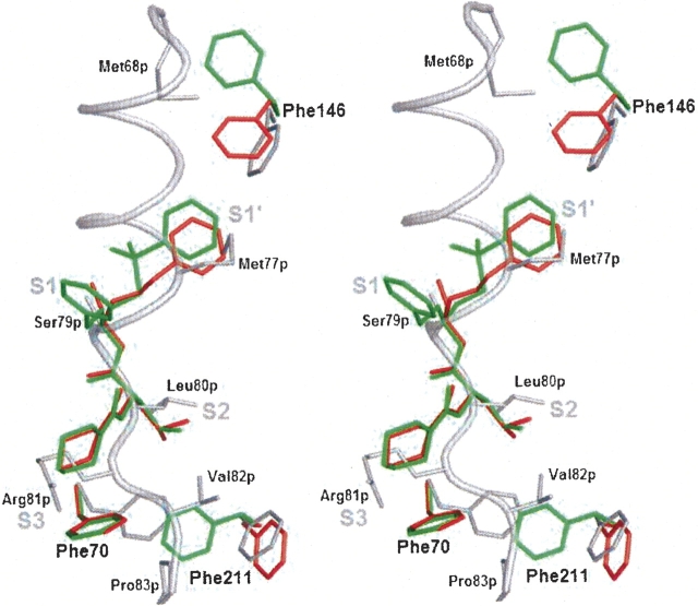
Figure 3.
Flexibility of Phe146, Phe70, and Phe211 lining the substrate binding cleft, illustrated by superposition of procathepsin S and two cathepsin S/inhibitor complexes. (Gray) procathepsin S (this work); (red) cathepsin S/morpholine-4-carboxylic acid {1S-(2-benzyloxy-1R-cyanoethylcarbamoyl)-3-methyl butyl} amide complex (PDB entry 1MS6); (green) cathepsin S/4-morpholinecarbonyl-Phe-(S-benzyl)Cys-ψ (CH=O) complex (PDB entry 1NPZ). Stereo picture of superposition prepared with PyMOL (DeLano 2002).

