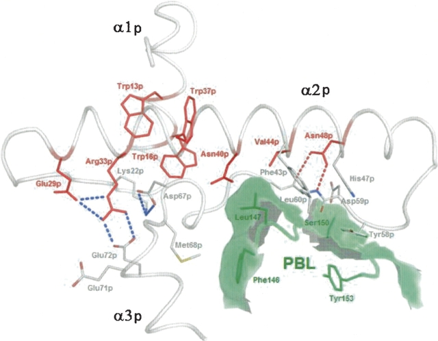
Figure 4.
Conserved residues involved in stabilization of the procathepsin S P-domain. Members of the ER(F/W)N(I/V)N motif are labeled in red. Dashed red lines symbolize hydrogen bonds, and blue lines symbolize salt bridges. Residues of the prosegment binding loop (PBL) of the enzyme are printed in dark green, the calculated contact area of PBL with the prosequence is shadowed in light green. Picture prepared with PyMOL (DeLano 2002).

